
Field Emission Scanning Electron Microscopes (FE-SEM)
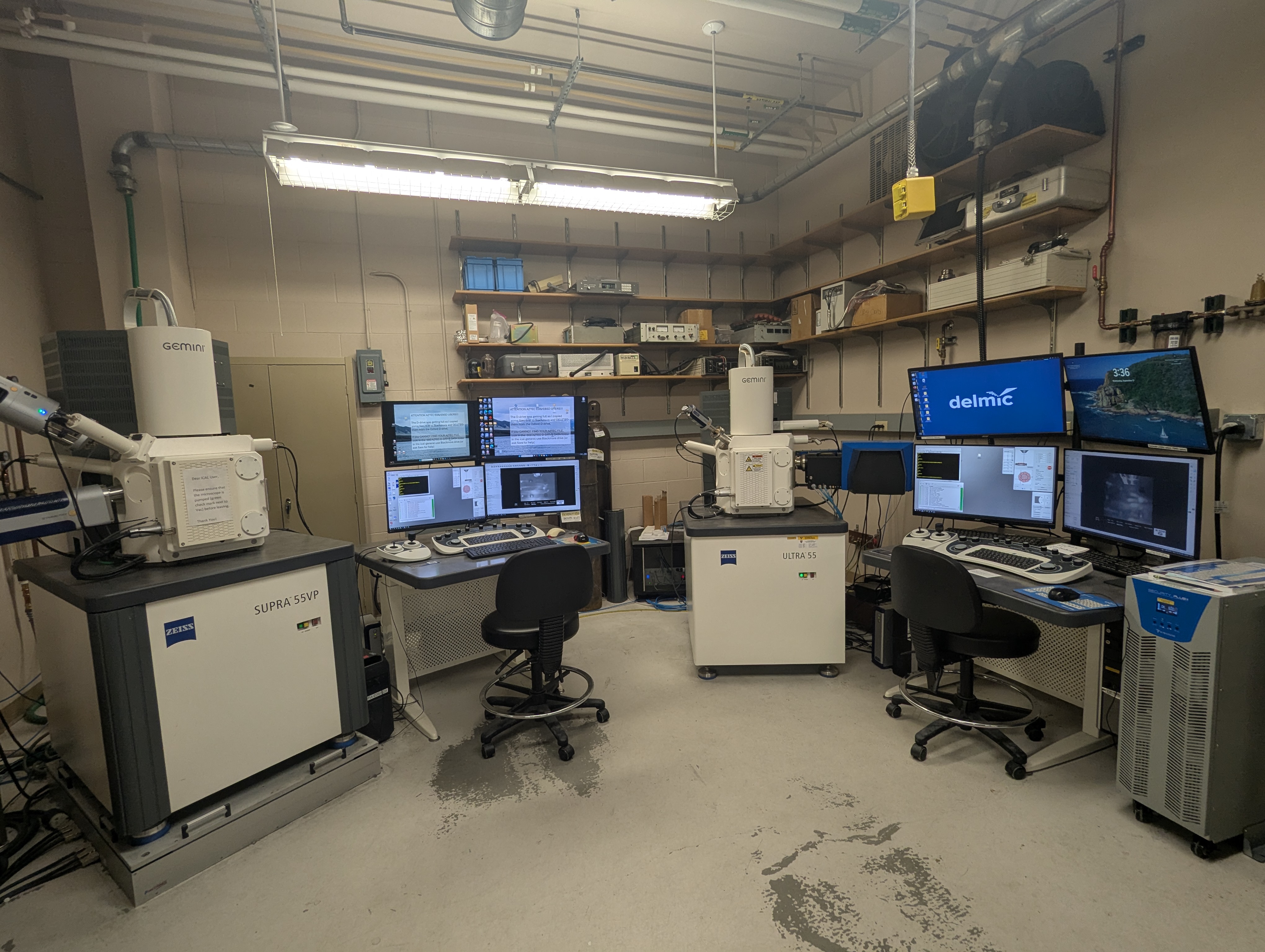
Zeiss SUPRA 55VP and ULTRA 55
Two Zeiss FE-SEMs provide powerful platforms for high-resolution (nanometer-scale) imaging and analysis methods used for characterization of materials (natural, engineered, biological). The two FE-SEMs have almost identical operating procedures, so the standard FE-SEM short course provides user access to both.
Capabilities of both instruments include:
- High-resolution imaging (down to 5 nanometers; using low beam voltage) of particle size, shape, distribution, texture using standard SEM imaging;
- Phase discrimination using back-scattered electron (BSE) imaging; materials with a large mean atomic number (Z) produce bright contrast compared with low Z materials (roughly correlated with density);
- Compositional analysis using Oxford Aztec energy dispersive spectroscopy (EDS), with data collected as spot (micron-scale) analyses and areal X-ray elemental mapping; semi-quantitative analyses are routinely obtainedand standards-based analyses can produce quantitative analyses. Detection limits of EDS are on the order of 0.1 wt%.
- Sample Requirements:
- Solid or biological materials can routinely be imaged or analyzed, but fluid or volatile materials should not be introduced into the vacuum sample chamber.
- Large volume sample analysis chamber allows large samples (cm-scale) to be imaged; but care must be taken regarding the height of samples to not impact the pole piece of the SEM, and large porous samples may take a long time to pump down in the vacuum chamber.
-
- Conducting materials may be used “as received”, but insulating materials should have a conducting coat applied to minimize charging problems. A metal coat (Au, Au/Pd, Ir) is typically used to optimize imaging whereas a C coat is typically used if elemental analysis (EDS) is emphasized.
- Critical point drying can be used as a final sample prep step for biological samples to mitigate drying artefacts. Typically, this step directly follows fixation/ethanol dehydration protocols. Samples may also be prepared using a cryo-microtome (available in the Center for Biofilm Engineering).
- Polished samples (thin sections, cut billets of metals, ceramics, rocks, fossils…) are best used for BSE imaging, and EDS point analyses or elemental X-ray maps. Particle mounts can be rapidly prepared by sprinkling particles onto conducting carbon “sticky dots”. Other irregular materials can be attached to metal sample holders but should not stand taller than ~1 cm.
Capabilities of the Zeiss Supra 55VP
- Oxford Electron Backscattered Diffraction (EBSD) for in situ phase identification based on diffraction patterns, determination of crystallographic preferred orientation of particles, and areal crystallographic orientation maps; EBSD and EDS detectors are integrated so phase identification using electron diffraction and elemental analysis using EDS can be obtained on the same areas of interest. For best results, ultra polishing with a Si-colloid suspension is typically required; this can be done in the Core Facility Polishing Lab. State-of-the-art Oxford Symmetry software allows rapid data reduction with search/match functions of diffraction data bases, stereo-net projections of crystallographic orientation and phase maps.
- Variable Pressure Mode: For insulating samples such as fossils or very rough surfaces that should not or cannot be adequately coated prior to SEM analysis, variable pressure mode can be used to prevent charging artefacts and allow secondary electron imaging analogue, backscattered electron imaging, and EDS/EBSD analysis. This mode is especially essential for Cryo-SEM, on ice samples or wet biological samples flash frozen and uncoatable. As a bonus, the detector used to create secondary electron image analogues is a visible light detector and can be used for pan-chromatic cathodoluminescence (CL) imaging during high-vacuum settings.
- Cryogenic SEM: a cryogenic stage is particularly useful for imaging biological materials and biofilms. Samples are frozen via immersion in liquid nitrogen, and this
- Tensile Stage: a tensile stage can be attached to the standard sample stage to observe deformation and brittle failure of materials under stress in real time.
Capabilities of the Ultra 55 FE-SEM
- Cathodoluminescence (CL) imaging: The Delmic CL detector is also equipped with a photospectrometer that detects luminescence from 400-900 nm. CL is widely used to characterize geologic materials (minerals, fossils, carbonate minerals, zircons and apatite for age dating) and in materials science (e.g., QA/QC determinations of heterogeneity of doped Si wafers, etc.). CL delivers compositional mapping based on distribution of trace element “activators” from the transition metals and Rare Earth elements. CL is used to obtain Panchromatic gray-scale or true color (using RGB filters) images to show variations in the distribution of trace elements at the ppm level. CL is sensitive to the chemical state of elements (e.g., Mn+2 vs Mn+ 3). The photospectrometer can be used to obtain spectral information on individual points to identify activator elements present, or conversely, signal can be obtained from a pre-defined limited range of wavelengths to map the distribution of an element of interest. Polished samples work best for CL imaging.
Please refer to these tutorial websites for further information on instruments and methods described above:
Electron Microbeam Methods