
What can ICAL do for you?
The Imaging and Chemical Analysis Laboratory (ICAL; RRID:SCR_026325) specializes in complementary surface, interfacial, and bulk characterization of materials using state-of-the-art instrumentation for high-resolution imaging and spectroscopy. ICAL is a user-oriented facility that conducts research in the physical, chemical, material, biological, Earth and environmental sciences, and allied engineering disciplines. ICAL supports fundamental and applied research done by academic users (student theses, faculty funded research), collaborators with colleagues from governmental agencies, as well as corporate clients to support technology transfer for economic growth in the region and nation.
ICAL is a proud partner of the Montana Nanotechnology Facility (MONT), one of 16 facilities in the United States in the NSF-sponsored (Grant# ECCS-2025391) National Nanotechnology Coordinated Infrastructure program (https://nnci.net/ ) which provides researchers from academia, industry, and government with access to cutting-edge fabrication and characterization tools, instrumentation, and expertise within all disciplines of nanoscale science, engineering, and technology. You can read more about MONT here.
Services
Our mission is to support great science and to develop great scientists (students, faculty, governmental and corporate partners). All potential users are welcome to access ICAL to support their research initiatives. ICAL services include:
- Training in the safe and effective use of modern characterization instrumentation. Short courses for each instrument are offered upon request to encourage users to conduct their own experiments. Once certified, users can schedule use of instruments as needed.
- Material Characterization: high resolution imaging and spectroscopic methods are available to document material morphologic features (size, shape, textures), crystal structure and crystallographic orientation, elemental composition (bulk), and surficial composition (atomic monolayers). We prefer that users be trained on the instruments and conduct their own services, with support from lab staff. However, samples may be submitted for characterization by lab staff to be done in consultation with clients.
- Expert Experimental Design and Data Interpretation: ICAL staff work closely with users and clients to define research questions to solve problems using the best possible procedures and protocols to produce meaningful results. This includes advice on sample selection, sample preparation, recommended instrumentation and operating parameters to optimize results, and data acquisition, reduction, and representation. We strive for rapid turn-around time from submission to research outcomes, and provide follow-on assistance to interpret data in appropriate contexts.
Instrumentation
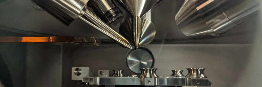
Instrumentation available at ICAL primarily includes electron-, Xray-, and particle-beam analytical instruments, that in aggregate provide complementary information on nano-scale to bulk chemical and physical properties of materials. These instruments include:
Two field-emissionscanning electron microscopes (FE-SEMs) for sub-micron imaging (optimally down to 5 nanometers spatial resolution); back-scattered electron (BSE) imaging is used for phase discrimination; Energy Dispersive Spectrometry (EDS) is used for fast X-ray elemental spot analyses and mapping, and standards-based quantitative analysis is possible. In addition:
- Electron Back-Scattered Diffraction (EBSD) is used for in situ phase identification using electron diffraction and for mapping of crystallographic preferred orientation of phases;
- Cathodoluminescence (CL) imaging is available to measure compositional heterogeneity of materials based on ppm-level variation of transition metal and REE abundances;
- A cryogenic stage is available for imaging delicate biological samples (e.g., biofilms)
- A tensile stage is available for conducting real-time deformation experiments in the FE-SEM.
For surface analysis of components (ions, molecular clusters, organic molecules) on material surfaces with a mass range of 1-10,000 amu, mass resolution of 10-3 amu, sub-micron spatial resolution, and detection limits in the ppm range.
Auger Nanoprobe, used for surficial elemental analysis (a few atomic monolayers on surfaces, ~1 nanometer); Auger Electron Spectroscopy (AES) can analyze all elements from Li and heavier, can provide quantitative analysis of elements on surfaces, and an ion sputter gun is used to provide elemental depth profiles across surfaces; In addition, this Auger Nanoprobe is uniquely equipped with:
- An EDS detector so the "bulk" composition can be compared with the surface composition; and,
- An EBSD detector, for phase identification and crystallographic orientation can be determined for the same analyzed area.
Used primarily for phase identification; Rietveld whole pattern fitting routines allow for quantitative analysis of samples; This instrument is also equipped with:
- Parallel Beam X-ray optics for a) Grazing Incident Diffraction (GID) of thin films, and b) Xray reflectometry (XRR) analysis for surface layer thickness, density, and roughness.
High-resolution atomic force microscope for nano-scale mapping of material surfaces; includes contact and tapping modes of operation, and numerous tips can be used to determine properties such as adhesion strength, magnetic force, mechanical properties, and can be functionalized to determine strength of chemical interactions.
For rapid major and trace element (ppm) analysis; can be used on metals, ceramics, powders, and for irregular materials such as art artifacts.
For determination of surface energy and hydrophilicity/hydrophobicity.
Obtains zeta potential values of nanoparticles in aqueous solution
- Carbon Coater for SEM sample preparation
- Metal Coater for SEM sample preparation (Au, Au/Pd, Ag, Ir)
- Critical Point Dryer for preparing biological samples for the FESEM
Please visit our instrumentation page for more complete descriptions of these instruments.
Tutorials and Technical Reports that Support Research at ICAL
Learn more about the instruments and methods the supports research in ICAL.
Research Activity in ICAL
Image Gallery - coming soon!
External Collaborations - coming soon!
Acknowledgment
Acknowledgment of core facility services and instruments supported by Montana State University is required in publications as it is a key performance indicator of our research activity and we are required to report publication output to our funding agencies. Learn more about how to acknowledge ICAL in your publication here.
Please report any recent publications to us by sending your DOI or a PDF using the link below: